POSTER
Stimulating 3D Skeletal Muscle Microtissues in a Novel Perfusable Microphysiological System with Integrated Electrodes
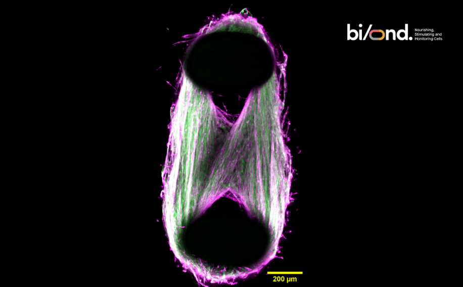
Find out how we have managed to successfully culture skeletal muscle cells (*) on our MUSbit™ platform to generate 3D skeletal muscle microtissues.
(*) The cells used for this study were bit.bio’s human iPSC-derived skeletal muscle cells (ioSkeletal Myocytes).
Download the poster now
If you want to:
- Form 3D muscle cell bundles on Bi/ond's MUSbit™ platform.
- Observe robust contractions triggered by electrical stimulation.
- Combine Bi/ond's cutting-edge technology with skeletal muscle bit.bio cells for measuring muscle tissue performance.
- Unlock research possibilities with MUSBit™/Let-it-Bit™ for pharmacological studies on wild-type and disease model cells.
This poster was originally presented by Dr. Mitchell Han, at the Microphysiological Systems (MPS) World Summit in Berlin, in June 2023.
Why does it matter?
The MUSbit™ is a perfusable organ-on-chip containing two pillars that serve as anchoring points for your muscle cell bundles. We have now gone one step further, integrating sensors (we call this new generation of chips Let-it-bit™).
Take your research to the next level by combining your tissue with a microfluidic channel! This integration allows you to study vascularization, dynamic drug response, and even mimic the immune system's response.
Furthermore, unlike other systems, the MUSbit ™ allows you to create 3D tissues with a limited number of cells, saving your budget without compromising the quality of your research.
In combination with integrated electrodes, you can pace and train your muscle tissue simulating real physiological conditions and achieve accelerated muscle maturation and enhanced cell stimulation, significantly advancing your research.

Our groundbreaking microphysiological system establishes a path towards creating more physiologically relevant biomimetic models for advancing drug development and disease modeling

.jpg)